LUMÉCEL
露美賽
Treatments
Lumécel 細胞治療
被時間磨鈍的身體自我修復力,由 Lumécel 重新喚醒。
What is Lumécel
什麼是 Lumécel 細胞治療?
Lumécel(露美賽)是 Etonne 整形外科診所的再生醫學細胞治療計畫。我們在自體血液細胞治療成分與脂肪來源再生成分之上,加入以光與溫度進行的 MCT 前處理,協助皮膚與組織具備得以修復的環境。
Who & Where
適合哪些人?
當您困擾於皮膚老化、發炎性或異位性皮膚問題、落髮與頭皮狀態,或希望一併照顧全身的恢復力時,可以考慮這項治療。
基本做法是直接輸送至臉部、頸部、手部、頭皮等需要的部位。若比起特定部位,您更在意整體狀態,則可考慮目的不同的靜脈注射,一併照顧全身。
我們優先考慮自體脂肪療程;若體型偏瘦,或因反覆施作而不易取得脂肪,則選擇自體血液療程。
| 適用對象與困擾 | 預期變化 | |
|---|---|---|
| Facial | 整體性的皮膚老化 | 改善皮膚狀態與膚質 |
| Skin | 異位性皮膚、發炎性皮膚問題、免疫相關皮膚問題等 | 改善皮膚狀態與膚質 |
| Hair | 落髮、頭皮狀態 | 改善毛髮密度與頭皮狀態 |
| IV injection | 全身狀態與恢復力的管理 | 以管理全身狀態為目的的使用 |
* 關於發炎性痤瘡與異位性皮膚的臨床證據,來自以自體血液細胞治療成分為對象的研究;毛髮密度則在自體血液成分與脂肪來源再生成分兩方面都有研究。24, 25, 26, 27, 28, 31, 32 反應與改善的程度因人而異,因此我們會先在諮詢中一起判斷這項治療現在是否適合您。靜脈注射是否適用,依檢查結果決定;所需的治療次數也因人而異。免疫相關的皮膚狀態,會先在諮詢中判斷是否可以適用。
Why Lumécel
重點不在用什麼,而在怎麼用
同樣是自體血液細胞治療成分與脂肪來源再生成分,處理方式不同,潛力也隨之不同。
Lumécel 細胞治療所使用的再生成分,其品質會因分離、濃縮與處理的方式而有很大差異。這正是 Etonne 在細胞處理系統上投注心力的原因。我們不只是把成分取出來,而是透過精密的分離與濃縮,以及穩定的溫度管理,營造對細胞負擔較小的條件。正如 Lumécel 這個名字所示,我們以光與溫度喚醒細胞。
閱讀更多收合
What We Use
再生醫學使用的是什麼?
我們使用的不是外來物質,而是來自您自己身體的自體來源成分。
COMPARISON
| 項目 | 脂肪幹細胞 | 自體血液細胞治療成分 |
|---|---|---|
| 來源 | 脂肪組織 | 血液 |
| 主要成分 | 多種再生細胞 | 血小板來源的生長因子與自體外泌體 |
| 運送的內容 | 細胞 | 訊號 |
| 核心角色 | 細胞定位後釋出訊號,營造修復環境 | 向您的細胞傳遞修復訊號 |
| 外泌體 | 由注入的細胞在體內分泌 | 自血小板取得並一併輸送 |
| 採集方式 | 以微創方式採集自體脂肪 | 抽取自體血液 |
閱讀更多收合
差異在於「運送的是什麼」。自體血液細胞治療成分運送的是訊號:血小板釋出的生長因子與自體外泌體,指示您的細胞進行修復。3, 4, 9 脂肪來源再生成分運送的是細胞:不只是幹細胞,還包含血管內皮細胞、血管周細胞、巨噬細胞的細胞群,在組織中安頓下來之後彼此交換訊號,並釋出生長因子與外泌體。11, 21, 22, 23 外泌體與其說是「內含」的,不如說是細胞在體內「製造」出來的。1, 3
在不易使用脂肪的情況下,血液是頗具吸引力的替代方案,但過去一直難以獲得符合期待的效果。然而,隨著相關技術在過去 20 年持續進步、證據逐漸累積,如今血液也已成為值得充分考慮的選項。18, 19 我們精密設計血漿純化的步驟,並同時施以波長經過精細調控的光與溫度前處理。
* 脂肪來源再生成分是未經培養、分離後直接使用的細胞混合物。細胞的組成比例因人與分離方式而異;動物與體外研究指出,比起單獨使用幹細胞,共存細胞之間的協同作用對效果有所貢獻。21, 22, 29, 30
How Does It Work
細胞治療是如何運作的?
再生細胞不是「磚塊」,而是「工地主任」。
效果何時開始出現?
細胞治療不像填充劑那樣帶來立即的變化。變化通常從約 1 週起,在 3~6 個月之間逐漸展現。
-
約 1 週
發炎逐漸平息
一開始進行的是整地的工作。療程引起的反應逐漸平息,修復得以開始的條件也隨之成形。發炎期會持續到這個時候,此時聚集的免疫細胞會清理受損部位,並釋出為下一階段做準備的訊號。注入的再生成分在這個時期也朝著傳遞修復訊號的方向發揮作用。
-
2~4 週
新血管逐漸增加
這是增生期的變化持續進行的階段。受損部位周圍長入新的微血管,氧氣與養分的供應增加,纖維母細胞聚集,開始建立新組織的基礎。比起外觀可見的變化,這是內部準備持續進行的時期。
-
1~3 個月
膠原蛋白增加
被激活的纖維母細胞會生成膠原蛋白和細胞外基質,使真皮層逐漸充盈飽滿。開始感受到膚質與彈性變化的時間點大多在此時,反應速度則因人而異。
-
3~6 個月
組織重新安定下來
這是重塑期。匆促生成的膠原蛋白轉變為更整齊的結構,暫時增加的血管只留下必要的部分,組織趨於穩定。由於這是變化定型的時期,是否需要追加療程,會依此時的狀態來判斷。
從診斷到施打,One-Stop
從診斷到施打,如何進行?
從診斷、抽血與取脂、細胞分離、前處理到施打,全程在 Etonne 之內以一條流程完成。
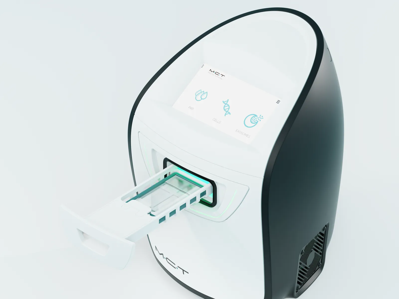
由整形外科專科醫師負責診斷與施打,並具備可減少組織損傷的取脂技術。Etonne 同時處理自體血液細胞治療成分與脂肪來源再生成分,並運作一套結合精密細胞分離與 MCT 前處理的處理系統。從抽血、取脂到再生治療一站式銜接,療程方案則依個人量身調整。
-
01
診斷
整形外科專科醫師確認皮膚狀態、病史與正在服用的藥物,並決定治療方法。掌握需要改善的部位之後,再決定以自體血液或脂肪之中的哪一種成分進行細胞治療。
-
02
抽血與取脂
抽血與取脂在隨時維持可進行手術狀態的空間中進行。空間的防疫與消毒、使用的器械,始終依該標準管理,並在此狀態下取得血液與脂肪。
-
03
細胞分離與前處理
細胞分離與純化所用的設備,是參考比較回收細胞數的資料後選定的。以這套設備完成分離與純化之後,再經過精細調校的 MCT 前處理,讓細胞準備好進入可以發揮作用的狀態。
-
04
施打
施打方式分為直接輸送至部位,以及透過靜脈注射輸送至全身兩種。依目的決定採用哪一種之後,由專科醫師對準所需的層次親自輸送。
* 有報告指出,脂肪來源成分的細胞分離所採用的拋棄式套組方式,比起人工操作,製程變異較小、回收量較穩定;我們團隊的內部測試也確認了同樣的趨勢。
我們設計的不是單一療程,而是修復的過程
再生醫學真正重要的,不是「哪種治療更好」,而是「什麼治療最適合我現在的狀態」。
Etonne 的 Lumécel 細胞治療,從準確診斷個人狀態開始,將合適的再生成分選擇、細胞分離與 MCT 前處理,到符合目的的輸送方式,設計為一條完整的流程。
一同拓展修復的範圍
- 協助術後快速恢復的方案
- 搭配提升皮膚狀態的肌膚活化注射(skin booster)
- 為求更好的改善,複合應用多種療程
閱讀更多收合
依需要單獨施作,或與其他療程合併進行,營造更有效的修復環境。
療程後如何照護?
療程隔天回診接受術後修復護理;若您接受的是自體脂肪療程,一週後再回診一次。
隔天
療程隔天請回診接受術後修復護理。兩種療程都會同時進行高壓氧治療。
閱讀更多收合
自體血液療程:以冷敷與舒緩護理讓臉部的施打部位平靜下來。護理將於當天完成。
自體脂肪療程:除了臉部的舒緩護理之外,也一併照護取脂的腹部。
一週後
若您接受的是自體脂肪療程,一週後請再回診一次拆線,並同時檢視臉部與腹部。自體血液療程沒有這次回診。這是因為自體脂肪療程需要照護的部位有兩處(施打部位與採集部位),而自體血液療程只有一處。
洗臉、化妝、飲酒、運動等日常生活中的注意事項,整理於下方的常見問題。
FAQ
我們整理了 Lumécel 細胞治療諮詢中常被問到的問題。
Lumécel 細胞治療對每個人的結果都一樣嗎?
細胞治療並非解決一切的萬能治療。反應與改善的程度有明顯的個人差異。不過,綜合至今的證據,Etonne 認為,在合適的對象上以正確的方法施作時,這是一項可以期待有意義改善的治療。
Lumécel 細胞治療安全嗎?
由於以自體血液與自體組織為基礎,只要遵守無菌原則施作,異物反應、過敏、感染的疑慮較低。不過,疑慮較低並不等於完全沒有反應。施打部位可能有輕微的腫脹或不適感,程度與持續時間因人而異。
Lumécel 細胞治療的疼痛程度如何?
多數人都能承受,不會感到勉強。成分濃度較高時可能感到些許痠脹,但不會持續太久。取脂時也只處理必要的範圍,因此療程後輕微的腫脹或不適感大約持續一兩天。自體脂肪療程因為採集部位也需要一併恢復,所需時間會較長。
Lumécel 細胞治療如何麻醉?
自體脂肪療程以舒眠麻醉進行。自體血液療程我們建議舒眠麻醉,但依狀態與療程範圍,也可以考慮局部麻醉。
Lumécel 細胞治療後會有瘀青或腫脹嗎?
可能出現輕微的瘀青或腫脹,但通常一兩天就會消退。自體脂肪療程在採集部位也可能有瘀青或不適感,消退所需的時間較長。
Lumécel 細胞治療後,何時可以恢復日常生活?
洗臉、化妝與運動從隔天起即可進行。不過,若採用的是自體脂肪療程,在取脂部位恢復的一週內,需要避免運動。飲酒建議一週後、三溫暖建議一個月後再開始。紫外線則建議在約三個月內盡量避免。依療程範圍與狀態,說明可能有所不同,我們會在療程當天再次向您確認。
Lumécel 細胞治療可以用於臉部以外的部位嗎?
頸部、手部、頭皮等需要再生的部位大致都可以應用。
Lumécel 細胞治療可以與其他療程合併進行嗎?
可以。可與多種療程合併進行,組合方式依目的與狀態決定。
參考文獻
在此說明本頁的內容是根據哪些文獻而撰寫的。
展開參考文獻收合參考文獻
以下文獻僅說明一般機轉,不保證治療效果。
- . Paracrine Mechanisms in Adult Stem Cell Signaling and Therapy. Circ Res. 2008. DOI 10.1161/CIRCRESAHA.108.176826 · PMID 19028920
- . Mesenchymal Stem Cells: Time to Change the Name! Stem Cells Transl Med. 2017. DOI 10.1002/sctm.17-0051 · PMID 28452204
- . Concise Review: MSC-Derived Exosomes for Cell-Free Therapy. Stem Cells. 2017. DOI 10.1002/stem.2575 · PMID 28294454
- . The biology, function, and biomedical applications of exosomes. Science. 2020. DOI 10.1126/science.aau6977 · PMID 32029601
- Growth factors and cytokines in wound healing. Wound Repair Regen. 2008. DOI 10.1111/j.1524-475X.2008.00410.x · PMID 19128254
- Exosomes released from human induced pluripotent stem cells-derived MSCs facilitate cutaneous wound healing by promoting collagen synthesis and angiogenesis. J Transl Med. 2015. DOI 10.1186/s12967-015-0417-0 · PMID 25638205
- Exosomes derived from human adipose mensenchymal stem cells accelerates cutaneous wound healing via optimizing the characteristics of fibroblasts. Sci Rep. 2016. DOI 10.1038/srep32993 · PMID 27615560
- Wound healing effect of adipose-derived stem cells: a critical role of secretory factors on human dermal fibroblasts. J Dermatol Sci. 2007. DOI 10.1016/j.jdermsci.2007.05.018 · PMID 17643966
- . A Review of Platelet-Rich Plasma: History, Biology, Mechanism of Action, and Classification. Skin Appendage Disord. 2018. DOI 10.1159/000477353 · PMID 29457008
- . From platelets to paracrine signals: A review of PRP, exosomes, and cell-based interventions for skin repair and rejuvenation. Nanomedicine. 2026. DOI 10.1016/j.nano.2026.102905 · PMID 41580060
- Stromal cells from the adipose tissue-derived stromal vascular fraction and culture expanded adipose tissue-derived stromal/stem cells: a joint statement of the International Federation for Adipose Therapeutics and Science (IFATS) and the International Society for Cellular Therapy (ISCT). Cytotherapy. 2013. DOI 10.1016/j.jcyt.2013.02.006 · PMID 23570660
- . Adipose tissue-derived stromal vascular fraction in regenerative medicine: a brief review on biology and translation. Stem Cell Res Ther. 2017. DOI 10.1186/s13287-017-0598-y · PMID 28619097
- . Photobiomodulation: A review of the molecular evidence for low level light therapy. J Plast Reconstr Aesthet Surg. 2021. DOI 10.1016/j.bjps.2020.12.059 · PMID 33436333
- . Proposed Mechanisms of Photobiomodulation or Low-Level Light Therapy. IEEE J Sel Top Quantum Electron. 2016. DOI 10.1109/JSTQE.2016.2561201 · PMID 28070154
- Low-level laser (light) therapy increases mitochondrial membrane potential and ATP synthesis in C2C12 myotubes with a peak response at 3-6 h. Photochem Photobiol. 2015. DOI 10.1111/php.12397 · PMID 25443662
- . Wound repair and regeneration: mechanisms, signaling, and translation. Sci Transl Med. 2014. DOI 10.1126/scitranslmed.3009337 · PMID 25473038
- . Angiogenesis in wound healing. J Investig Dermatol Symp Proc. 2000. DOI 10.1046/j.1087-0024.2000.00014.x · PMID 11147674
- Efficacy and safety of photothermal biomodulated platelet-rich plasma versus standard platelet-rich plasma for facial rejuvenation: a prospective split-face randomized study. Sci Rep. 2026. DOI 10.1038/s41598-026-54310-9 · PMID 42225690
- Photothermal Biostimulation of Platelet-Rich Plasma Improves Hand Rejuvenation Clinical Outcome: A Pilot Study. Photobiomodul Photomed Laser Surg. 2024. DOI 10.1089/pho.2023.0192 · PMID 38836757
- . The Effect of Photobiomodulation on Human Mesenchymal Cells: A Literature Review. Aesthetic Plast Surg. 2021. DOI 10.1007/s00266-021-02173-y · PMID 33616715
- Characterization of freshly isolated and cultured cells derived from the fatty and fluid portions of liposuction aspirates. J Cell Physiol. 2006. DOI 10.1002/jcp.20636 · PMID 16557516
- . The Adipose Stromal Vascular Fraction as a Complex Cellular Source for Tissue Engineering Applications. Tissue Eng Part B Rev. 2017. DOI 10.1089/ten.TEB.2017.0061 · PMID 28316259
- The ratio of ADSCs to HSC-progenitors in adipose tissue derived SVF may provide the key to predict the outcome of stem-cell therapy. Clin Transl Med. 2018. DOI 10.1186/s40169-018-0183-8 · PMID 29417261
- . Clinical evaluation of efficacy of intralesional platelet-rich plasma injection versus 1064 nm long-pulsed Neodymium:YAG laser in the treatment of inflammatory acne vulgaris in adolescent and post-adolescent patients: a prospective randomized split-face comparative study. Lasers Med Sci. 2022. DOI 10.1007/s10103-022-03510-6 · PMID 35084634
- Platelet rich plasma injection versus topical erythromycin 2% in treatment of acne vulgaris. J Dermatolog Treat. 2020. DOI 10.1080/09546634.2020.1793884 · PMID 32643473
- . Possible clinical efficacy and tolerability of platelet-rich plasma with atopic dermatitis. J Cosmet Dermatol. 2021. DOI 10.1111/jocd.14170 · PMID 34114728
- Platelet-Rich Plasma for Androgenetic Alopecia: A Systematic Review and Meta-Analysis of Randomized Controlled Trials. J Cutan Med Surg. 2023. DOI 10.1177/12034754231191461 · PMID 37533146
- Platelet-rich plasma for the treatment of alopecia: a systematic review and meta-analysis. Blood Transfus. 2021. DOI 10.2450/2021.0216-21 · PMID 34967722
- Human uncultured adipose-derived stromal vascular fraction shows therapeutic potential against osteoarthritis in immunodeficient rats via direct effects of transplanted M2 macrophages. Stem Cell Res Ther. 2024. DOI 10.1186/s13287-024-03946-3 · PMID 39334434
- Paracrine effect of the stromal vascular fraction containing M2 macrophages on human chondrocytes through the Smad2/3 signaling pathway. J Cell Physiol. 2022. DOI 10.1002/jcp.30823 · PMID 35766589
- The safety and efficacy of autologous adipose-derived stromal vascular fraction for nonscarring alopecia: A systematic review. Arch Dermatol Res. 2021. DOI 10.1007/s00403-021-02238-7 · PMID 34014340
- . Efficacy of autologous stromal vascular fraction injection in the treatment of androgenic alopecia. Arch Dermatol Res. 2022. DOI 10.1007/s00403-022-02501-5 · PMID 36525057
- . Wound repair and regeneration. Eur Surg Res. 2012. DOI 10.1159/000339613 · PMID 22797712
- . Cellular and molecular mechanisms of skin wound healing. Nat Rev Mol Cell Biol. 2024. DOI 10.1038/s41580-024-00715-1 · PMID 38528155
Gnecchi M, Zhang Z, Ni A, Dzau VJ. Paracrine Mechanisms in Adult Stem Cell Signaling and Therapy. Circ Res. 2008.
Caplan AI. Mesenchymal Stem Cells: Time to Change the Name! Stem Cells Transl Med. 2017.
Phinney DG, Pittenger MF. Concise Review: MSC-Derived Exosomes for Cell-Free Therapy. Stem Cells. 2017.
Kalluri R, LeBleu VS. The biology, function, and biomedical applications of exosomes. Science. 2020.
Barrientos S, Stojadinovic O, Golinko MS, et al. Growth factors and cytokines in wound healing. Wound Repair Regen. 2008.
Zhang J, Guan J, Niu X, et al. Exosomes released from human induced pluripotent stem cells-derived MSCs facilitate cutaneous wound healing by promoting collagen synthesis and angiogenesis. J Transl Med. 2015.
Hu L, Wang J, Zhou X, et al. Exosomes derived from human adipose mensenchymal stem cells accelerates cutaneous wound healing via optimizing the characteristics of fibroblasts. Sci Rep. 2016.
Kim WS, Park BS, Sung JH, et al. Wound healing effect of adipose-derived stem cells: a critical role of secretory factors on human dermal fibroblasts. J Dermatol Sci. 2007.
Alves R, Grimalt R. A Review of Platelet-Rich Plasma: History, Biology, Mechanism of Action, and Classification. Skin Appendage Disord. 2018.
Esmaeili E, Rad I. From platelets to paracrine signals: A review of PRP, exosomes, and cell-based interventions for skin repair and rejuvenation. Nanomedicine. 2026.
Bourin P, Bunnell BA, Casteilla L, et al. Stromal cells from the adipose tissue-derived stromal vascular fraction and culture expanded adipose tissue-derived stromal/stem cells: a joint statement of the International Federation for Adipose Therapeutics and Science (IFATS) and the International Society for Cellular Therapy (ISCT). Cytotherapy. 2013.
Bora P, Majumdar AS. Adipose tissue-derived stromal vascular fraction in regenerative medicine: a brief review on biology and translation. Stem Cell Res Ther. 2017.
Glass GE. Photobiomodulation: A review of the molecular evidence for low level light therapy. J Plast Reconstr Aesthet Surg. 2021.
de Freitas LF, Hamblin MR. Proposed Mechanisms of Photobiomodulation or Low-Level Light Therapy. IEEE J Sel Top Quantum Electron. 2016.
Ferraresi C, Kaippert B, Avci P, et al. Low-level laser (light) therapy increases mitochondrial membrane potential and ATP synthesis in C2C12 myotubes with a peak response at 3-6 h. Photochem Photobiol. 2015.
Eming SA, Martin P, Tomic-Canic M. Wound repair and regeneration: mechanisms, signaling, and translation. Sci Transl Med. 2014.
Tonnesen MG, Feng X, Clark RA. Angiogenesis in wound healing. J Investig Dermatol Symp Proc. 2000.
Wanitphakdeedecha R, Ollero MPGCR, Bhorntarakcharoen W, et al. Efficacy and safety of photothermal biomodulated platelet-rich plasma versus standard platelet-rich plasma for facial rejuvenation: a prospective split-face randomized study. Sci Rep. 2026.
Tejero García P, Mota Antigua S, Ortega Zamorano M, et al. Photothermal Biostimulation of Platelet-Rich Plasma Improves Hand Rejuvenation Clinical Outcome: A Pilot Study. Photobiomodul Photomed Laser Surg. 2024.
Pinto H, Goñi Oliver P, Sánchez-Vizcaíno Mengual E. The Effect of Photobiomodulation on Human Mesenchymal Cells: A Literature Review. Aesthetic Plast Surg. 2021.
Yoshimura K, Shigeura T, Matsumoto D, et al. Characterization of freshly isolated and cultured cells derived from the fatty and fluid portions of liposuction aspirates. J Cell Physiol. 2006.
Ramakrishnan VM, Boyd NL. The Adipose Stromal Vascular Fraction as a Complex Cellular Source for Tissue Engineering Applications. Tissue Eng Part B Rev. 2017.
Kilinc MO, Santidrian A, Minev I, et al. The ratio of ADSCs to HSC-progenitors in adipose tissue derived SVF may provide the key to predict the outcome of stem-cell therapy. Clin Transl Med. 2018.
Moftah NH, Mansour AM, Ibrahim SMA. Clinical evaluation of efficacy of intralesional platelet-rich plasma injection versus 1064 nm long-pulsed Neodymium:YAG laser in the treatment of inflammatory acne vulgaris in adolescent and post-adolescent patients: a prospective randomized split-face comparative study. Lasers Med Sci. 2022.
Ibrahim ZA, Gheida SF, El-Halaby AR, et al. Platelet rich plasma injection versus topical erythromycin 2% in treatment of acne vulgaris. J Dermatolog Treat. 2020.
Yosef A, Elkady N, Khattab F. Possible clinical efficacy and tolerability of platelet-rich plasma with atopic dermatitis. J Cosmet Dermatol. 2021.
Zhang X, Ji Y, Zhou M, et al. Platelet-Rich Plasma for Androgenetic Alopecia: A Systematic Review and Meta-Analysis of Randomized Controlled Trials. J Cutan Med Surg. 2023.
Cruciani M, Masiello F, Pati I, et al. Platelet-rich plasma for the treatment of alopecia: a systematic review and meta-analysis. Blood Transfus. 2021.
Onoi Y, Matsumoto T, Anjiki K, et al. Human uncultured adipose-derived stromal vascular fraction shows therapeutic potential against osteoarthritis in immunodeficient rats via direct effects of transplanted M2 macrophages. Stem Cell Res Ther. 2024.
Fujita M, Matsumoto T, Hayashi S, et al. Paracrine effect of the stromal vascular fraction containing M2 macrophages on human chondrocytes through the Smad2/3 signaling pathway. J Cell Physiol. 2022.
Kang BY, Li AW, Lee MH, et al. The safety and efficacy of autologous adipose-derived stromal vascular fraction for nonscarring alopecia: A systematic review. Arch Dermatol Res. 2021.
El-Khalawany M, Rageh MA, Elnokrashy I, Ibrahim SMA. Efficacy of autologous stromal vascular fraction injection in the treatment of androgenic alopecia. Arch Dermatol Res. 2022.
Reinke JM, Sorg H. Wound repair and regeneration. Eur Surg Res. 2012.
Peña OA, Martin P. Cellular and molecular mechanisms of skin wound healing. Nat Rev Mol Cell Biol. 2024.

醫學審閱:金學永, 曺延穆 院長 · 最後審閱