LUMÉCEL
Lumécel
Treatments
Lumécel cell therapy
Time dulls the body's own capacity to heal. Lumécel awakens it again.

What is Lumécel
What is Lumécel cell therapy?
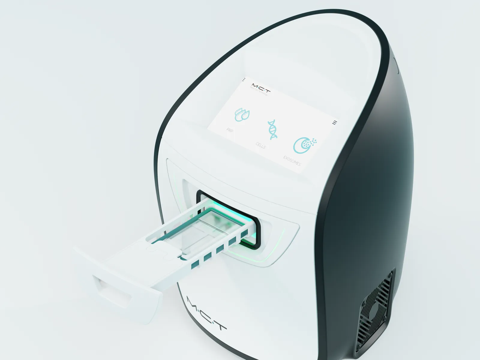
Lumécel is the regenerative medicine cell therapy program of Etonne Plastic Surgery. It combines an autologous blood-derived cell therapy component and an adipose-derived regenerative component with MCT preconditioning using light and temperature, helping your skin and tissue build the conditions they need to recover.
Who & Where
Who can it help?
It is a treatment to consider when skin aging, inflammatory and atopic skin conditions, or hair loss and scalp condition are a concern, and when you also want to care for the recovery capacity of the whole body.
The standard approach delivers the treatment directly to the area that needs it, such as the face, neck, hands or scalp. When your overall condition is a greater concern than any one area, we consider an intravenous infusion, which serves a different purpose and addresses the whole body alongside it.
We consider a fat-based treatment first. When your build is slim, or when repeated treatments have made fat difficult to obtain, we choose a blood-based treatment instead.
| For whom · Concerns | Expected change | |
|---|---|---|
| Facial | General skin aging | Improved skin condition and texture |
| Skin | Atopic skin, inflammatory skin conditions, immune-related skin conditions and more | Improved skin condition and texture |
| Hair | Hair loss, scalp condition | Improved hair density and scalp condition |
| IV injection | Overall condition and recovery capacity | Used to support overall condition |
* The clinical evidence on inflammatory acne and atopic skin comes from studies of the blood-derived component; for hair density, there are studies of both the blood-derived and the fat-derived components.24, 25, 26, 27, 28, 31, 32 Response and degree of improvement vary from person to person, so we first assess in consultation whether this treatment is right for you now. Whether an intravenous infusion is suitable is decided from test results, and the number of treatments needed also varies. For immune-related skin conditions, we assess first whether it can be applied.
Why Lumécel
Not what we use, but how we use it
The same autologous blood-derived cell therapy component and adipose-derived regenerative component hold different potential depending on how they are handled.
The quality of the regenerative components used in Lumécel cell therapy can vary greatly with how they are isolated, concentrated and processed. This is why Etonne invests so much care in its cell processing system. Rather than simply extracting the components, we create conditions that place less stress on the cells, through precise isolation and concentration and stable temperature control. True to the name Lumécel, we awaken cells with light and temperature.
Read moreShow less
What we pay attention to
- Autologous exosomes. Exosomes are the carriers of recovery signals that cells release. In a blood-based treatment, we obtain them from your own platelets and deliver them.3, 4
- Multiple growth factors. Growth factors involved in skin regeneration, including VEGF, EGF, TGF, FGF-2 and PDGF, act together.5, 10
- Increased cellular energy (ATP). We support the energy metabolism that cells need to recover.15
- Light and temperature control (PBM). We keep light and temperature conditions constant to stimulate the cells.13, 14, 20
* The degree of response varies from person to person. The equipment used is the MCT Unit and MCT Kit from Metacell Technology. For cell isolation of the fat-derived component, we use SmartX, a kit-based cell isolation device, in which isolation and washing take place inside a single-use kit.
What We Use
What does regenerative medicine use?
Not foreign substances, but autologous components that come from your own body.
COMPARISON
| Category | Adipose-derived stem cells | Autologous blood-derived cell therapy component |
|---|---|---|
| Origin | Fat tissue | Blood |
| Main components | Multiple regenerative cells | Platelet-derived growth factors and autologous exosomes |
| What is carried | Cells | Signals |
| Core role | Cells settle in and send signals to create a recovery environment | Delivers recovery signals to your cells |
| Exosomes | Secreted inside the body by the injected cells | Obtained from platelets and delivered together |
| How it is collected | Minimally invasive fat harvest | A simple blood draw |
Lumécel cell therapy does not inject foreign substances. It draws on autologous components that already exist in your body. The two main ones are the autologous blood-derived cell therapy component and the adipose-derived stem cell therapy component (SVF/ADSC, hereafter "adipose-derived regenerative component"). Both come from your own body, but they differ in composition and in how they act. The blood-derived component mainly delivers recovery signals, whereas the fat-derived component directly supplies the cells that take part in regeneration.8, 9, 11, 12
Read moreShow less
The difference lies in what is carried. What the blood-derived component carries is signals: the growth factors and autologous exosomes released by platelets instruct your cells to recover.3, 4, 9 What the fat-derived component carries is cells. Not stem cells alone, but a mixed population that also includes endothelial cells, pericytes and macrophages settles in, exchanges signals, and releases growth factors and exosomes.11, 21, 22, 23 Exosomes are less something that is "contained" than something the cells "produce" inside the body.1, 3
When fat is difficult to use, blood is an attractive alternative, though for a long time it was hard to obtain the results people hoped for. Over the past 20 years, however, the related technology has steadily advanced and evidence has accumulated, and blood has now become an option well worth considering.18, 19 We design the plasma purification step with precision and apply light and temperature preconditioning with finely tuned wavelengths alongside it.
* The adipose-derived regenerative component is a mixture of cells isolated and used immediately without culture. The proportions of cell types differ by person and isolation method, and animal and in vitro studies report that the cooperation of the accompanying cells contributes to the effect, compared with using stem cells alone.21, 22, 29, 30
How Does It Work
How does cell therapy work?
Regenerative cells are not the "bricks" but the "site foreman."
In Lumécel cell therapy, the injected regenerative cells act by sending recovery signals to surrounding cells rather than by becoming tissue themselves.1, 2 In the past, the explanation was that "the injected stem cells turn directly into young skin cells or blood vessel cells." But research to date is converging on the view that this is not the main mechanism. Rather than surviving for long, the injected cells release a range of signaling molecules over days to weeks and set the surrounding cells in motion.1, 2
So regenerative cells are less like "bricks" that build tissue directly and more like a "site foreman" directing the entire construction. Instead of making tissue themselves, they instruct the surrounding cells:
- "Reduce inflammation"
- "Make collagen"
- "Form new blood vessels"
- "Wake up the dormant stem cells"
What these signals do
Together, these signals work to calm chronic inflammation, promote tissue regeneration including collagen and blood vessels, and awaken your body's own dormant stem cells.3, 10
Read moreShow less
What happens in the skin
Applied to the skin, the process generally unfolds in this order: growth factor release → fibroblast activation → formation of new blood vessels → increase in collagen and elastin → increase in skin thickness and firmness → reduction in fine lines.6, 7
Exosomes
Recently, exosomes have drawn attention as the key to this process. Exosomes are tiny carriers released by cells; they hold gene-regulating material (microRNA), proteins and growth factors, and pass recovery signals on to other cells.4 Broadly speaking, it is accurate to think of regenerative cells as "factories that produce exosomes."3
This is why Etonne does not say "they turn into young cells." "The signals your own body sends out reawaken its capacity to regenerate." This explanation better matches the current scientific evidence, and it is how Etonne explains the treatment to patients.
When do the effects appear?
Cell therapy does not create an immediate change the way fillers do. Change usually begins around 1 week and continues gradually over 3–6 months.
-
Around 1 week
Inflammation settles
At first, the ground is prepared. The reaction caused by the treatment subsides, and the conditions for recovery to begin are created. The inflammatory phase continues up to about this point, and the immune cells that gather during it clear the damaged area and send out signals that prepare the next stage. The injected regenerative components also act during this period by delivering recovery signals.
-
2–4 weeks
New blood vessels increase
This is the period when the changes of the proliferative phase take place. New capillaries grow in around the damaged area, increasing the supply of oxygen and nutrients, and fibroblasts gather and begin to lay the foundation for new tissue. It is a time when preparation proceeds inside rather than showing on the surface.
-
1–3 months
Collagen increases
Activated fibroblasts produce collagen and extracellular matrix, and the dermis fills in. This is generally when you begin to notice changes in skin texture and firmness, though the speed of response varies from person to person.
-
3–6 months
The tissue settles into place
This is the remodeling phase. The collagen that was produced quickly is reorganized into a more orderly structure, only as many of the temporarily increased blood vessels as are needed remain, and the tissue stabilizes. Because this is when the change settles, whether further treatment is needed is decided by looking at your condition around this time.
Read moreShow less
* The periods above are approximate divisions based on Etonne's clinical practice and vary with your condition, the extent of the treatment, and whether it was blood-based or fat-based. The order of the four stages follows the general course of skin recovery (inflammatory → proliferative → remodeling phase).
From diagnosis to treatment, One-Stop
How does it proceed, from diagnosis to treatment?
From diagnosis through blood and fat collection, cell isolation, preconditioning and treatment, everything flows as one process within Etonne.
Board-certified plastic surgeons handle both diagnosis and treatment, with fat harvesting techniques that reduce tissue damage. Etonne works with both the autologous blood-derived cell therapy component and the adipose-derived regenerative component, and operates a processing system that combines precise cell isolation with MCT preconditioning. From blood and fat collection to regenerative treatment, everything is one-stop, and the protocol is tailored to each individual.
-
01
Diagnosis
A board-certified plastic surgeon reviews your skin condition, medical history and current medications, and decides on the treatment approach. After identifying the areas that need improvement, the surgeon determines whether to proceed with cell therapy using the blood-derived or the fat-derived component.
-
02
Blood and fat collection
Blood and fat are collected in a space maintained at all times in a condition ready for surgery. The disinfection and sterilization of the space and the instruments we use are always kept to that standard, and blood and fat are obtained under those conditions.
-
03
Cell isolation and preconditioning
The equipment we use for cell isolation and purification was chosen after reviewing data comparing the number of cells recovered. Once isolation and purification are complete with this equipment, the cells go through precisely adjusted MCT preconditioning and are prepared to do their work.
-
04
Treatment
The treatment is delivered either directly to the area or to the whole body by intravenous infusion. Once the route is chosen according to the purpose, the surgeon delivers it personally, matching the layer that needs it.
* The single-use kit method used for cell isolation of the fat-derived component has been reported to show smaller process variation and more consistent yields than manual handling, and our team's internal testing has confirmed the same tendency.
Not a single procedure, but a designed process of recovery
What truly matters in regenerative medicine is not "which treatment is better" but "which treatment best fits my condition right now."
Etonne designs Lumécel cell therapy as one continuous process. It begins with an accurate diagnosis of your individual condition and continues through the choice of a suitable regenerative component, cell isolation and MCT preconditioning, and a delivery method that fits the purpose.
Broadening the scope of recovery together
- Solutions that support faster recovery after surgery
- Skin booster treatments combined to lift skin condition
- Combined application of several procedures for better improvement
Read moreShow less
Depending on your needs, it is applied on its own or combined with other treatments to create a more effective environment for recovery.
How is aftercare handled?
You receive recovery care at the clinic the day after treatment, and if you had a fat-based treatment, you visit once more a week later.
The next day
You come to the clinic the day after treatment for recovery care. Both treatments include hyperbaric oxygen therapy.
Read moreShow less
Blood-based treatment. Cooling and soothing care calm the treated areas of the face. Care is completed on this one day.
Fat-based treatment. In addition to soothing care for the face, the abdomen where fat was harvested is cared for as well.
One week later
If you had a fat-based treatment, you come once more a week later for suture removal and a check of both the face and the abdomen. A blood-based treatment does not include this visit. This is because a fat-based treatment has two areas to care for (the treated area and the donor site), while a blood-based treatment has one.
Everyday precautions such as washing your face, makeup, alcohol and exercise are covered in the frequently asked questions below.
FAQ
Questions we are often asked during consultations about Lumécel cell therapy.
Does Lumécel cell therapy give everyone the same results?
Cell therapy is not a cure-all that solves everything. Response and degree of improvement clearly vary from person to person. That said, taking the evidence to date as a whole, Etonne's judgment is that it is a treatment from which meaningful improvement can be expected when performed in the right way for the right candidates.
Is Lumécel cell therapy safe?
Because it is based on your own blood and your own tissue, the risk of foreign-body reaction, allergic reaction and infection is low when the treatment is performed with sterile technique. However, a low risk is not the same as no reaction at all. Mild swelling or discomfort at the treated area can occur, and its degree and duration vary from person to person.
How much does Lumécel cell therapy hurt?
For most people it is easily tolerable. When the component is highly concentrated you may feel a dull, heavy sensation, but it does not last long. Fat harvesting is also limited to the area needed, so mild swelling or discomfort after the treatment typically lasts about a day or two. A fat-based treatment takes longer, as the donor site recovers at the same time.
What kind of anesthesia is used for Lumécel cell therapy?
A fat-based treatment is performed under sedation. For a blood-based treatment we recommend sedation, but local anesthesia can also be considered depending on your condition and the extent of the treatment.
Will there be bruising or swelling after Lumécel cell therapy?
Slight bruising or swelling can occur, but it usually subsides within a day or two. With a fat-based treatment, the donor site may also show bruising or discomfort, and it takes longer to settle.
When can I return to daily life after Lumécel cell therapy?
Washing your face, makeup and exercise are possible from the next day. If you had a fat-based treatment, however, you should refrain from exercise for one week while the donor site recovers. We recommend no alcohol for one week and no sauna for one month. We also recommend avoiding sun exposure as much as possible for about three months. Guidance may differ with the extent of the treatment and your condition, so we confirm it again on the day of treatment.
Can Lumécel cell therapy be used on areas other than the face?
It can be applied to any area that needs regeneration, including the neck, hands and scalp.
Can Lumécel cell therapy be combined with other treatments?
Yes. It can be combined with a range of treatments, and the combination is decided according to your goals and condition.
References
The literature on which the explanations on this page are based.
Show the full reference listHide the reference list
These references describe general mechanisms and do not guarantee treatment outcomes.
- . Paracrine Mechanisms in Adult Stem Cell Signaling and Therapy. Circ Res. 2008. DOI 10.1161/CIRCRESAHA.108.176826 · PMID 19028920
- . Mesenchymal Stem Cells: Time to Change the Name! Stem Cells Transl Med. 2017. DOI 10.1002/sctm.17-0051 · PMID 28452204
- . Concise Review: MSC-Derived Exosomes for Cell-Free Therapy. Stem Cells. 2017. DOI 10.1002/stem.2575 · PMID 28294454
- . The biology, function, and biomedical applications of exosomes. Science. 2020. DOI 10.1126/science.aau6977 · PMID 32029601
- Growth factors and cytokines in wound healing. Wound Repair Regen. 2008. DOI 10.1111/j.1524-475X.2008.00410.x · PMID 19128254
- Exosomes released from human induced pluripotent stem cells-derived MSCs facilitate cutaneous wound healing by promoting collagen synthesis and angiogenesis. J Transl Med. 2015. DOI 10.1186/s12967-015-0417-0 · PMID 25638205
- Exosomes derived from human adipose mensenchymal stem cells accelerates cutaneous wound healing via optimizing the characteristics of fibroblasts. Sci Rep. 2016. DOI 10.1038/srep32993 · PMID 27615560
- Wound healing effect of adipose-derived stem cells: a critical role of secretory factors on human dermal fibroblasts. J Dermatol Sci. 2007. DOI 10.1016/j.jdermsci.2007.05.018 · PMID 17643966
- . A Review of Platelet-Rich Plasma: History, Biology, Mechanism of Action, and Classification. Skin Appendage Disord. 2018. DOI 10.1159/000477353 · PMID 29457008
- . From platelets to paracrine signals: A review of PRP, exosomes, and cell-based interventions for skin repair and rejuvenation. Nanomedicine. 2026. DOI 10.1016/j.nano.2026.102905 · PMID 41580060
- Stromal cells from the adipose tissue-derived stromal vascular fraction and culture expanded adipose tissue-derived stromal/stem cells: a joint statement of the International Federation for Adipose Therapeutics and Science (IFATS) and the International Society for Cellular Therapy (ISCT). Cytotherapy. 2013. DOI 10.1016/j.jcyt.2013.02.006 · PMID 23570660
- . Adipose tissue-derived stromal vascular fraction in regenerative medicine: a brief review on biology and translation. Stem Cell Res Ther. 2017. DOI 10.1186/s13287-017-0598-y · PMID 28619097
- . Photobiomodulation: A review of the molecular evidence for low level light therapy. J Plast Reconstr Aesthet Surg. 2021. DOI 10.1016/j.bjps.2020.12.059 · PMID 33436333
- . Proposed Mechanisms of Photobiomodulation or Low-Level Light Therapy. IEEE J Sel Top Quantum Electron. 2016. DOI 10.1109/JSTQE.2016.2561201 · PMID 28070154
- Low-level laser (light) therapy increases mitochondrial membrane potential and ATP synthesis in C2C12 myotubes with a peak response at 3-6 h. Photochem Photobiol. 2015. DOI 10.1111/php.12397 · PMID 25443662
- . Wound repair and regeneration: mechanisms, signaling, and translation. Sci Transl Med. 2014. DOI 10.1126/scitranslmed.3009337 · PMID 25473038
- . Angiogenesis in wound healing. J Investig Dermatol Symp Proc. 2000. DOI 10.1046/j.1087-0024.2000.00014.x · PMID 11147674
- Efficacy and safety of photothermal biomodulated platelet-rich plasma versus standard platelet-rich plasma for facial rejuvenation: a prospective split-face randomized study. Sci Rep. 2026. DOI 10.1038/s41598-026-54310-9 · PMID 42225690
- Photothermal Biostimulation of Platelet-Rich Plasma Improves Hand Rejuvenation Clinical Outcome: A Pilot Study. Photobiomodul Photomed Laser Surg. 2024. DOI 10.1089/pho.2023.0192 · PMID 38836757
- . The Effect of Photobiomodulation on Human Mesenchymal Cells: A Literature Review. Aesthetic Plast Surg. 2021. DOI 10.1007/s00266-021-02173-y · PMID 33616715
- Characterization of freshly isolated and cultured cells derived from the fatty and fluid portions of liposuction aspirates. J Cell Physiol. 2006. DOI 10.1002/jcp.20636 · PMID 16557516
- . The Adipose Stromal Vascular Fraction as a Complex Cellular Source for Tissue Engineering Applications. Tissue Eng Part B Rev. 2017. DOI 10.1089/ten.TEB.2017.0061 · PMID 28316259
- The ratio of ADSCs to HSC-progenitors in adipose tissue derived SVF may provide the key to predict the outcome of stem-cell therapy. Clin Transl Med. 2018. DOI 10.1186/s40169-018-0183-8 · PMID 29417261
- . Clinical evaluation of efficacy of intralesional platelet-rich plasma injection versus 1064 nm long-pulsed Neodymium:YAG laser in the treatment of inflammatory acne vulgaris in adolescent and post-adolescent patients: a prospective randomized split-face comparative study. Lasers Med Sci. 2022. DOI 10.1007/s10103-022-03510-6 · PMID 35084634
- Platelet rich plasma injection versus topical erythromycin 2% in treatment of acne vulgaris. J Dermatolog Treat. 2020. DOI 10.1080/09546634.2020.1793884 · PMID 32643473
- . Possible clinical efficacy and tolerability of platelet-rich plasma with atopic dermatitis. J Cosmet Dermatol. 2021. DOI 10.1111/jocd.14170 · PMID 34114728
- Platelet-Rich Plasma for Androgenetic Alopecia: A Systematic Review and Meta-Analysis of Randomized Controlled Trials. J Cutan Med Surg. 2023. DOI 10.1177/12034754231191461 · PMID 37533146
- Platelet-rich plasma for the treatment of alopecia: a systematic review and meta-analysis. Blood Transfus. 2021. DOI 10.2450/2021.0216-21 · PMID 34967722
- Human uncultured adipose-derived stromal vascular fraction shows therapeutic potential against osteoarthritis in immunodeficient rats via direct effects of transplanted M2 macrophages. Stem Cell Res Ther. 2024. DOI 10.1186/s13287-024-03946-3 · PMID 39334434
- Paracrine effect of the stromal vascular fraction containing M2 macrophages on human chondrocytes through the Smad2/3 signaling pathway. J Cell Physiol. 2022. DOI 10.1002/jcp.30823 · PMID 35766589
- The safety and efficacy of autologous adipose-derived stromal vascular fraction for nonscarring alopecia: A systematic review. Arch Dermatol Res. 2021. DOI 10.1007/s00403-021-02238-7 · PMID 34014340
- . Efficacy of autologous stromal vascular fraction injection in the treatment of androgenic alopecia. Arch Dermatol Res. 2022. DOI 10.1007/s00403-022-02501-5 · PMID 36525057
- . Wound repair and regeneration. Eur Surg Res. 2012. DOI 10.1159/000339613 · PMID 22797712
- . Cellular and molecular mechanisms of skin wound healing. Nat Rev Mol Cell Biol. 2024. DOI 10.1038/s41580-024-00715-1 · PMID 38528155
Gnecchi M, Zhang Z, Ni A, Dzau VJ. Paracrine Mechanisms in Adult Stem Cell Signaling and Therapy. Circ Res. 2008.
Caplan AI. Mesenchymal Stem Cells: Time to Change the Name! Stem Cells Transl Med. 2017.
Phinney DG, Pittenger MF. Concise Review: MSC-Derived Exosomes for Cell-Free Therapy. Stem Cells. 2017.
Kalluri R, LeBleu VS. The biology, function, and biomedical applications of exosomes. Science. 2020.
Barrientos S, Stojadinovic O, Golinko MS, et al. Growth factors and cytokines in wound healing. Wound Repair Regen. 2008.
Zhang J, Guan J, Niu X, et al. Exosomes released from human induced pluripotent stem cells-derived MSCs facilitate cutaneous wound healing by promoting collagen synthesis and angiogenesis. J Transl Med. 2015.
Hu L, Wang J, Zhou X, et al. Exosomes derived from human adipose mensenchymal stem cells accelerates cutaneous wound healing via optimizing the characteristics of fibroblasts. Sci Rep. 2016.
Kim WS, Park BS, Sung JH, et al. Wound healing effect of adipose-derived stem cells: a critical role of secretory factors on human dermal fibroblasts. J Dermatol Sci. 2007.
Alves R, Grimalt R. A Review of Platelet-Rich Plasma: History, Biology, Mechanism of Action, and Classification. Skin Appendage Disord. 2018.
Esmaeili E, Rad I. From platelets to paracrine signals: A review of PRP, exosomes, and cell-based interventions for skin repair and rejuvenation. Nanomedicine. 2026.
Bourin P, Bunnell BA, Casteilla L, et al. Stromal cells from the adipose tissue-derived stromal vascular fraction and culture expanded adipose tissue-derived stromal/stem cells: a joint statement of the International Federation for Adipose Therapeutics and Science (IFATS) and the International Society for Cellular Therapy (ISCT). Cytotherapy. 2013.
Bora P, Majumdar AS. Adipose tissue-derived stromal vascular fraction in regenerative medicine: a brief review on biology and translation. Stem Cell Res Ther. 2017.
Glass GE. Photobiomodulation: A review of the molecular evidence for low level light therapy. J Plast Reconstr Aesthet Surg. 2021.
de Freitas LF, Hamblin MR. Proposed Mechanisms of Photobiomodulation or Low-Level Light Therapy. IEEE J Sel Top Quantum Electron. 2016.
Ferraresi C, Kaippert B, Avci P, et al. Low-level laser (light) therapy increases mitochondrial membrane potential and ATP synthesis in C2C12 myotubes with a peak response at 3-6 h. Photochem Photobiol. 2015.
Eming SA, Martin P, Tomic-Canic M. Wound repair and regeneration: mechanisms, signaling, and translation. Sci Transl Med. 2014.
Tonnesen MG, Feng X, Clark RA. Angiogenesis in wound healing. J Investig Dermatol Symp Proc. 2000.
Wanitphakdeedecha R, Ollero MPGCR, Bhorntarakcharoen W, et al. Efficacy and safety of photothermal biomodulated platelet-rich plasma versus standard platelet-rich plasma for facial rejuvenation: a prospective split-face randomized study. Sci Rep. 2026.
Tejero García P, Mota Antigua S, Ortega Zamorano M, et al. Photothermal Biostimulation of Platelet-Rich Plasma Improves Hand Rejuvenation Clinical Outcome: A Pilot Study. Photobiomodul Photomed Laser Surg. 2024.
Pinto H, Goñi Oliver P, Sánchez-Vizcaíno Mengual E. The Effect of Photobiomodulation on Human Mesenchymal Cells: A Literature Review. Aesthetic Plast Surg. 2021.
Yoshimura K, Shigeura T, Matsumoto D, et al. Characterization of freshly isolated and cultured cells derived from the fatty and fluid portions of liposuction aspirates. J Cell Physiol. 2006.
Ramakrishnan VM, Boyd NL. The Adipose Stromal Vascular Fraction as a Complex Cellular Source for Tissue Engineering Applications. Tissue Eng Part B Rev. 2017.
Kilinc MO, Santidrian A, Minev I, et al. The ratio of ADSCs to HSC-progenitors in adipose tissue derived SVF may provide the key to predict the outcome of stem-cell therapy. Clin Transl Med. 2018.
Moftah NH, Mansour AM, Ibrahim SMA. Clinical evaluation of efficacy of intralesional platelet-rich plasma injection versus 1064 nm long-pulsed Neodymium:YAG laser in the treatment of inflammatory acne vulgaris in adolescent and post-adolescent patients: a prospective randomized split-face comparative study. Lasers Med Sci. 2022.
Ibrahim ZA, Gheida SF, El-Halaby AR, et al. Platelet rich plasma injection versus topical erythromycin 2% in treatment of acne vulgaris. J Dermatolog Treat. 2020.
Yosef A, Elkady N, Khattab F. Possible clinical efficacy and tolerability of platelet-rich plasma with atopic dermatitis. J Cosmet Dermatol. 2021.
Zhang X, Ji Y, Zhou M, et al. Platelet-Rich Plasma for Androgenetic Alopecia: A Systematic Review and Meta-Analysis of Randomized Controlled Trials. J Cutan Med Surg. 2023.
Cruciani M, Masiello F, Pati I, et al. Platelet-rich plasma for the treatment of alopecia: a systematic review and meta-analysis. Blood Transfus. 2021.
Onoi Y, Matsumoto T, Anjiki K, et al. Human uncultured adipose-derived stromal vascular fraction shows therapeutic potential against osteoarthritis in immunodeficient rats via direct effects of transplanted M2 macrophages. Stem Cell Res Ther. 2024.
Fujita M, Matsumoto T, Hayashi S, et al. Paracrine effect of the stromal vascular fraction containing M2 macrophages on human chondrocytes through the Smad2/3 signaling pathway. J Cell Physiol. 2022.
Kang BY, Li AW, Lee MH, et al. The safety and efficacy of autologous adipose-derived stromal vascular fraction for nonscarring alopecia: A systematic review. Arch Dermatol Res. 2021.
El-Khalawany M, Rageh MA, Elnokrashy I, Ibrahim SMA. Efficacy of autologous stromal vascular fraction injection in the treatment of androgenic alopecia. Arch Dermatol Res. 2022.
Reinke JM, Sorg H. Wound repair and regeneration. Eur Surg Res. 2012.
Peña OA, Martin P. Cellular and molecular mechanisms of skin wound healing. Nat Rev Mol Cell Biol. 2024.
We start by asking whether this treatment is right for you now
Before recommending any treatment, Etonne first assesses with you whether this treatment is right for you at this time.
Medical review: Kim Harkyoung, Cho Jeongmok (Director) · Last reviewed